


Department of Nuclear Medicine
Our service
Presentation of the department
The Nuclear Medicine Department of Princess Grace Hospital carries out conventional scans (known as SPECT scans) and positron emission tomography (PET scans). It has a 64-slice PET scanner, two SPECT cameras (including one with a scanner) and will soon have a specialised CZT semi-conductor detector for cardiology.
Conventional scans
The most common scans include bone scintigraphy to investigate rheumatological and infectious diseases or cancer, cardiac perfusion imaging, either to investigate heart disease or to investigate heart rhythm or left ventricular function, lung scans to investigate pulmonary embolism or for assessment prior to surgery, neuroendocrine tumour scans (somatostatin receptors, MIBG), brain scans in Parkinson’s disease, thyroid scans, lymphoscintigraphy for sentinel lymph node mapping and in lymphoedema of the limbs, and renal scans.
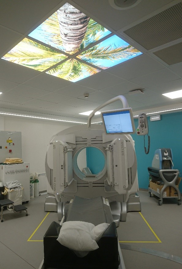
GE NM/CT 870 CZT
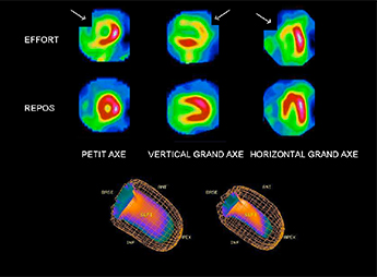
Scintigraphie myocardique
First two lines: stress images are shown on the top line with the images at rest on the second line. Myocardial perfusion imaging has shown an area that is poorly perfused under stress in the anterior wall of the left ventricle, as well as the apex and septum (in orange on the image, as shown by the arrows), and normal at rest (red), thus suggesting myocardial ischaemia.
The line at the bottom of the image shows the three-dimensional representation in cinema mode. Global heart contractility is normal.

Example of bone scintigraphy focusing on a compression fracture of the 4th lumbar vertebra.
Positron emission tomography (PET)
Several kinds of PET scans can be performed, depending on the tracer used. The most common scan, which is carried out every day, is the 18F-FDG (glucose derivative) PET scan. However, it requires preparation in that the patient must strictly fast for 6 hours (contact the department for more information). This examination is prescribed in cancer cases, as well as for infectious/inflammatory or neurological conditions.
Other tracers may be used, but need to be specially ordered and delivered. These include 18F-Choline (prostate cancer), 18F-DOPA (gastrointestinal carcinoid tumours), and very soon 18F-Florbetapir (Alzheimer’s disease), 18FNa (cancer and rheumatological investigation of the skeleton) and 18F-Oestradiol (investigation of oestrogen receptors of breast tumours).
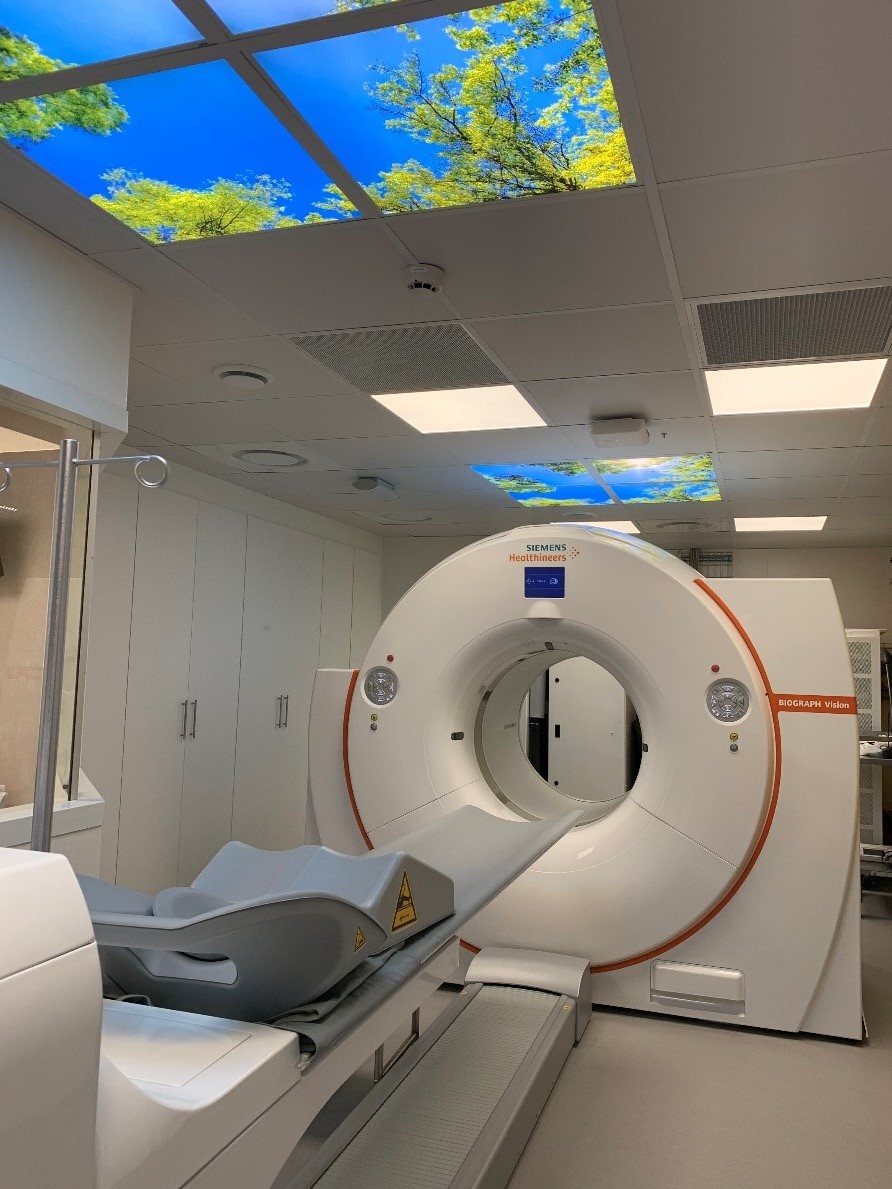
TEP digital SIEMENS Vision à très haute résolution

Illustration of a PET scan requested for lung cancer: this examination shows a cancer nodule on the right lung (yellow arrows) and an affected lymph node (blue arrow), with no other abnormalities suggestive of distant metastases, meaning that the patient is eligible for surgery.
This patient also has inflammatory osteoarthritis of the left shoulder and a dilated upper urinary tract on the left (black arrows).

Illustration of a brain PET scan: a 74-year-old patient with mild temporal and spatial disorientation. The examination shows that some regions of the brain (dark blue areas) have diminished metabolic activity compared with the same regions in normal subjects. This examination points to incipient Alzheimer’s disease.

